Diese Seite steht derzeit nicht in Ihrer Sprache zur Verfügung. Mittels der
Übersetzungsfunktion
von Google kann Ihnen eine automatische Übersetzung angezeigt werden. Bitte beachten Sie jedoch, dass wir keinerlei Verantwortung für diese
Dienstleistung tragen und die Übersetzung auch nicht von uns geprüft wurde.
Wenn Sie weitere Unterstützung benötigen,
kontaktieren Sie uns bitte.
Raman spectroscopy for oncology research (and drug delivery research)
March 2014
Raman microscopy is routinely used for characterisation and identification of material, but the need for this molecular imaging and analysis technique has become increasingly important in biology.
Renishaw Inc (USA) recently contributed to a webinar about Raman spectroscopy for oncology research (and drug delivery research). This proved very popular, with attendees learning about:
- a Raman technique used to identify cancer cells from healthy cells
- a Raman technique used to analyse lipid based drug discovery
- set up of an experiment using label-free detection (no fluorescent dyes, colorimetric stains or labelled antibodies needed)
The webinar is available to view on Lab Manager's website.
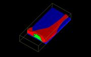
Image: Multiple component 3D Raman volume image of glioma cell. This shows the substrate (blue), cell (red) and nucleus (green). Renishaw thanks Dr Matthew Baker, University of Central Lancashire, for providing the cell sample.
News updates
Register for regular news updates from Renishaw