Biowissenschaften
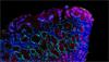
Die Raman-Spektroskopie wird erfolgreich zur Analyse von menschlichen, tierischen und pflanzlichen Zellen und Geweben eingesetzt.
- Bioprocessing
- Strukturanalyse von Proteinen/Peptiden
- Mikrobiologie
- Drug delivery in vitro und in vivo
- Krebsforschung/Pathologie
- Redoxbiologie
- Regenerative Medizin
- Alterungsprozesse und neurodegenerative Erkrankungen,
- Biokraftstoff und Agrarforschung
- Lipidomics
- Metabolomics
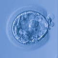
- Entwicklungsbiologie
- Reproduktionsbiologie
- Virologie
Klicken Sie auf die folgenden Links, um zu erfahren, wie wir Ihnen mit Ihren Anwendungen im Bereich der Biowissenschaften behilflich sein können:


Wir sind für Sie da
Mehr Informationen über diesen Anwendungsbereich, bzw. einer Anwendung, die hier nicht angesprochen wird, erhalten Sie von unserem Anwendungsteam.
Kontaktieren Sie unser AnwendungsteamWebinar – Resonanz-Raman-Spektroskopie für die redoxbiologische Forschung
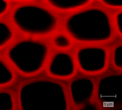
Die Resonanz-Raman-Spektroskopie (RR-Spektroskopie) ist die ideale Technik für die redoxbiologische Forschung. Sie reagiert nicht nur hochempfindlich auf Hämproteine, sondern kann auch Aufschluss über ihre Oxidation und Oxygenierungsgrade in situ (Lösung, Organellen, Zellen und Gewebe) geben. RR Imaging bietet sowohl chemische als auch räumliche Daten, durch die sich Korrelationen zwischen Hämproteinverteilung, Sauerstoffsättigung und Protein-/Zellfunktionen herstellen lassen.
Webinar anschauenDownloads: Biowissenschaften
-
 Brochure: Biological analysis using Raman spectroscopy and imaging [en]
The domain of biological research is shaped by our ability to peer into the world of the small. Simply seeing microscopic biological samples is useful, but by utilising Raman spectroscopy we can surpass sight into the molecular realm… and beyond! Download this brochure to discover the wealth of biological applications made possible by Renishaw's Raman systems.
-
 Anwendungshinweis: Redoxbiologie mit dem inVia konfokalem Raman-Mikroskop [it]
Die Raman-Spektroskopie reagiert empfindlich auf das Vorhandensein von Hämproteinen und eignet sich ideal für die Untersuchung ihrer Redoxbiologie, ohne eine Isolation bzw. Färbung. Das Redox der Hämproteine ist eng mit ihren Proteinfunktionen verbunden - Transport und Speicherung von Sauerstoff, Elektronentransport und Auffangen freier Radikaler. Durch den Einsatz der Raman-Spektroskopie, zur Beschreibung von Redox-Zuständen innerhalb biologischer Systeme, können Forscher die Redox-Dynamik und seine Auswirkungen auf Gesundheitsvorschriften und Krankheiten untersuchen.
-
 Application note: Raman imaging for biological applications. No stains. No labels. [en]
Raman spectroscopy is an information-rich, label-free, non-invasive imaging technique that is ideal for life sciences research. It uses laser light scattering to provide a chemical fingerprint at each point of the analysed area and identifies the molecules present in samples.
-
 Product note: Microplate mapping with Renishaw Raman system's [en]
Renishaw’s microplate mapping package enables researchers to use Renishaw’s Raman spectroscopy products to rapidly and easily analyse material contained in microplates.


-
Raman chemical imaging for life sciences [en]
Short movie to demonstrate the benefits of using the inVia confocal Raman microscope for powerful, flexible, Raman Imaging for life sciences applications.